Skip to content

From the American College of Rheumatology

Rheumatology for Primary Care
  • Symptoms
  • Diseases
    • ANCA-Associated Vasculitis
    • Behcet’s Syndrome
    • Chronic Recurrent Multifocal Osteomyelitis
    • Crystalline Arthropathies
    • Drug or Medication-induced Rheumatic Diseases
    • Giant Cell Arteritis
    • Henoch-Schönlein Purpura (HSP)
    • IgG4-Related Disease
    • Juvenile Dermatomyositis
    • Juvenile Idiopathic Arthritis
    • Kawasaki Disease
    • Mixed Connective Tissue Disease (MCTD)
    • Multisystem Inflammatory Syndrome in Children (MIS-C)
    • Myositis
    • Polyarteritis Nodosa
    • Polymyalgia Rheumatica
    • Rheumatoid Arthritis (RA)
    • Sarcoidosis
    • Sicca/Sjogren’s Disease
    • Spondyloarthritis
    • Systemic Lupus Erythematosus (SLE)
    • Systemic Sclerosis
  • Diagnostics
  • Case Studies
    • ANCA-Associated Vasculitis
    • Behcet’s Syndrome
    • Crystalline Arthropathies
    • Giant Cell Arteritis
    • Henoch-Schönlein Purpura
    • IgG4-Related Disease
    • Juvenile Dermatomyositis
    • Juvenile Inflammatory Arthritis
    • Kawasaki Disease
    • Mixed Connective Tissue Disease
    • Polyarteritis Nodosa
    • Polymalgia Rheumatica
    • Rheumatoid Arthritis
    • Sarcoidosis
    • Sicca/Sjogren’s Disease
    • Spondyloarthritis
    • Systemic Lupus Erythematosus
    • Systemic Sclerosis
  • Meds
    • Treatment Guides
    • Addressing Patient Fears
    • Treatment Considerations
    • Major Categories of Rheumatology Medications
  • Resources
    • Vaccination Considerations – Pediatrics
    • Vaccination Considerations – Adults

Mixed Connective Tissue Disease Case Studies

Home » Mixed Connective Tissue Disease Case Studies
Mixed Connective Tissue Disease Case Studies
  • Case 1

  • Case 2

  • Case 1

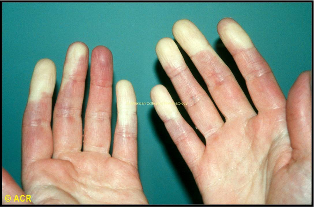
A 24 yo F with no PMH with 6 months of joint pain and puffy hands. Mother with systemic lupus erythematosus. ROS notable for Raynaud’s (view image) and mild dyspnea on exertion.

Physical Exam

  • Synovitis of MCPs and wrists
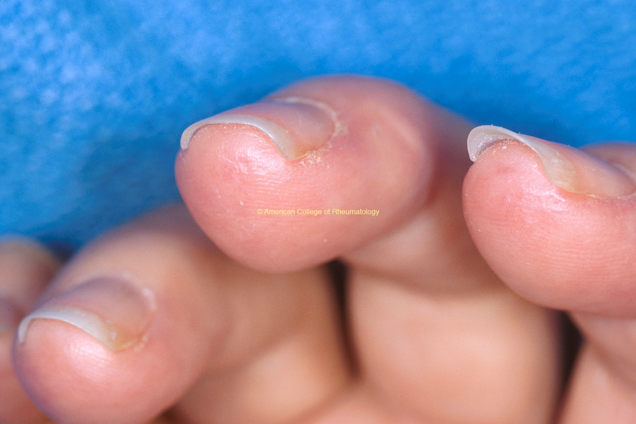
  • Pitting at distal fingertips (view image); nailfold capillaroscopy
  • Normal pulmonary exam; RRR, prominent S2

Diagnostic Workup

  • Labs:
    • Hemoglobin 10.6, MCV 82; remaining CBC and BMP WNL
    • High titer ANA, speckled pattern; high titer RNP; Negative Smith, Ro, La, dsDNA, centromere, Scl 70, RF, CCP antibodies
  • Imaging:
    • CXR: Normal lung fields
    • TTE: Dilated RV with normal function; normal LV size and function

Treatment & Further Workup

  • Hydroxychloroquine for inflammatory arthritis
  • Calcium channel blocker for Raynaud’s
  • Right heart catheterization for workup of pulmonary artery hypertension
  • Case 2

A 29 yo F presents to the ED for acute-onset, electric shock pain over the L side of the face. Diagnosed with trigeminal neuralgia. ROS also notable for trouble going up and down stairs and dyspnea on exertion.

Physical Exam

  • Mildly puffy/swollen hands; no synovitis
  • Severe tenderness to palpation of L face
  • 4-/5 strength of BL iliopsoas and deltoids; otherwise normal neurologic exam
  • Normal cardiopulmonary exam
  • No rashes

Diagnostic Workup

  • Labs:
    • High titer ANA, speckled pattern; high titer RNP; Negative Smith, Ro, La, dsDNA, centromere, Scl 70, RF, CCP antibodies, myositis panel
    • CK 1,037
  • Imaging:
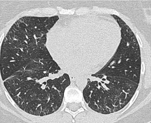
    • CXR shows reticular opacities at the BL bases (view image); CT demonstrates NSIP pattern of ILD (view image); TTE normal
    • MRI thigh with diffuse muscle edema (view image)

Treatment

  • Prednisone 40mg daily
  • Carbamazepine for trigeminal neuralgia
  • Mycophenolate mofetil for ILD and myositis

Raynaud's Phenomenon: Hands

Raynaud's Phenomenon: Hands

The well demarcated pallor present in the fingers of both hands is characteristic of Raynaud’s phenomenon- a manifestation of ischemia from peripheral vasospasm. Raynaud’s is common in limited scleroderma, diffuse scleroderma, and MCTD but may also occur in the absence of any underlying disease.

Raynaud's Phenomenon: Hands

Digital pitting scars on hands

Digital pitting scars on the pulps of the index and middle fingers, secondary to chronic microvascular disease, are depicted in a patient with complaints of Raynaud’s

Chest x-ray showing interstitial markings

Chest radiograph showing diffuse reticular interstitial markings

Chest CT Showing Interstitial Markings

CT imaging of chest showing patchy consolidation in both lower lobes and increased interstitial markings in the periphery of the lungs

Thigh MRI

STIR magnetic resonance image of the proximal thigh muscles; inflammation appears as bright areas

View Information About Mixed Connective Tissue Disease
Tami Bonnett-Admi2024-02-14T23:14:22+00:00

What do you think about this website?

We would love to hear your feedback.
Provide Feedback

Rheumatology Directory

Access this ACR resource to locate licensed rheumatology providers in your area for referrals.
Find A Rheumatology Provider

ACR Information

About ACR

ACR Contacts

Website Feedback

What do you think about the website? We would love to hear your feedback.

Provide Feedback

Authors

This product was funded by a medical grant from Pfizer. The content was written by a physician work group. See Authors.

© Copyright American College of Rheumatology 2026 | All rights reserved | See ACR Policies

Page load link
Go to Top